Why Are Medical Tubes Used in Surgery? This question reaches beyond simple equipment. During an operation, tubes may deliver oxygen, remove fluid, administer medication, drain blood, or monitor pressure. Some are visible beside the patient’s abdomen. Others remain inside the airway, bladder, or blood vessel.
So, what is the role of medical tubes in surgery? Their central purpose is controlled access. A tube creates a predictable pathway when direct access is unsafe or impractical. Endotracheal tubes support ventilation. Suction tubes clear blood and secretions. Catheters measure urine output and help clinicians assess kidney function. Surgical drains reduce fluid accumulation, although their value depends on careful selection and monitoring.
The World Health Organization’s Global Patient Safety Report 2024 states that approximately one in ten patients experiences harm in healthcare, with more than half considered preventable. Tubes can improve safety, but they can also introduce infection, blockage, tissue injury, or accidental removal. That tension matters. A tube is never harmless by default.
Sir Berkeley Moynihan, a respected British surgeon, is often attributed with saying, “A drainage tube is a confession of surgical failure.” The statement is deliberately provocative, not universally correct. Modern surgery uses drains for specific clinical reasons, not emotional reassurance. Fortune Business Insights’ 2024 Medical Tubing Market report also reflects sustained demand for specialized medical tubing, driven partly by minimally invasive procedures.
This article examines tube functions, materials, placement, and risks. It also asks a practical question: when does a tube protect the patient, and when might it create avoidable complexity?
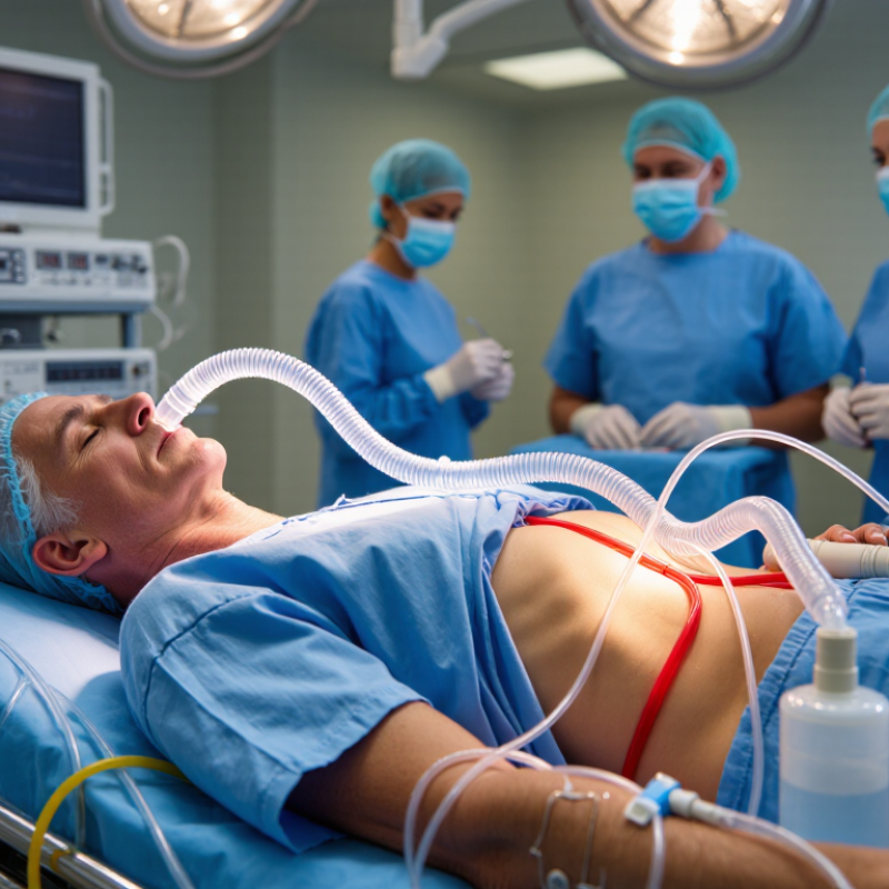
Medical tubes are narrow, sterile devices that move air, fluids, medicine, or waste during surgery. An endotracheal tube passes through the mouth into the windpipe, helping the anesthesia team control breathing. A urinary catheter drains the bladder. A nasogastric tube can remove stomach contents after abdominal procedures. Surgical drains carry blood or fluid away from an incision into a collection bulb.
Their purpose is practical, not decorative. A tube may keep a patient’s airway open, reduce pressure inside the stomach, or prevent fluid from collecting near delicate tissue. Small markings help clinicians check insertion depth. A soft cuff can seal the airway. A visible drainage line lets staff measure output at the bedside.
The need is substantial. The World Health Organization has estimated that more than 234 million major operations occur globally each year. AHRQ reports that urinary catheters are used in roughly 15% to 25% of hospitalized patients. However, tubes are not harmless. CDC guidance links unnecessary urinary-catheter use with catheter-associated urinary tract infections, so clinicians should remove them promptly when they are no longer needed. That decision can be imperfect. A tube may protect one system while irritating another. Careful placement, daily review, and clean handling remain essential.
Why Are Medical Tubes Used in Surgery?
During surgery, anesthesia can relax the muscles that keep the airway open. It can also reduce natural breathing. An anesthesiologist may place a breathing tube through the mouth and into the windpipe. This tube connects to a ventilator, which delivers controlled amounts of oxygen and anesthetic gases. The team watches oxygen levels, carbon dioxide, heart rate, and blood pressure continuously. The goal is simple. Keep breathing safe.
Not every patient needs one. Some procedures use a face mask or a smaller airway device instead. The choice depends on the operation, anesthesia depth, body position, and medical history. A small inflatable cuff may seal the breathing tube and reduce leakage. Suction equipment can remove saliva or fluids when needed. These details seem routine, but placement requires skill and constant checking. No tube is completely risk-free. A sore throat, hoarse voice, or mild irritation can occur afterward. Serious problems are uncommon, yet preparation matters.
Tips: Tell the anesthesia team about asthma, sleep apnea, loose teeth, allergies, or past airway problems. Do not hide recent coughs or breathing changes. Follow fasting instructions carefully. Ask what type of airway support is planned, although the final decision may change during surgery. It is reasonable to feel nervous. Clear questions often make the process less mysterious. Some explanations may sound technical, and patients may need to ask twice. That is okay.
| Tube or Device | Where It Is Placed | Primary Function | How It Supports Anesthesia or Breathing | Typical Use Considerations | Key Safety Monitoring |
|---|---|---|---|---|---|
| Endotracheal Tube | Through the mouth or nose into the trachea | Maintains an open airway and provides a sealed route for ventilation | Allows an anesthesia machine or ventilator to deliver oxygen and anesthetic gases while helping remove carbon dioxide | Commonly used during general anesthesia, especially when controlled ventilation or airway protection is needed | Position confirmation, oxygen level, carbon dioxide level, airway pressure, and cuff pressure |
| Supraglottic Airway | Above the vocal cords, typically over the entrance to the larynx | Creates an airway without passing through the vocal cords | Provides a connection for spontaneous or assisted ventilation and delivery of anesthetic gases | Often suitable for selected short procedures in patients with an appropriate risk of aspiration | Airway seal, ventilation quality, oxygen saturation, and signs of obstruction or leakage |
| Nasal Cannula | Small prongs positioned inside the nostrils | Delivers supplemental oxygen | Supports oxygenation during monitored anesthesia care, recovery, or procedures performed without a secured airway | Useful when the patient is breathing independently and only low-to-moderate oxygen support is required | Breathing effort, oxygen saturation, respiratory rate, and carbon dioxide monitoring when indicated |
| Face Mask | Over the nose and mouth | Delivers oxygen and can assist ventilation with a manual resuscitation bag | Provides short-term breathing support during induction of anesthesia, emergence, or urgent airway management | Usually used as a temporary method because it does not provide the same airway protection as a tracheal tube | Mask seal, chest movement, oxygen saturation, carbon dioxide level, and gastric inflation risk |
| Breathing Circuit | Connects the airway device to the anesthesia machine | Carries oxygen and anesthetic gases to the patient and returns exhaled gases | Enables delivery of a controlled gas mixture and supports manual or mechanical ventilation | Selected according to the patient, procedure, ventilation requirements, and anesthesia equipment | Connections, leaks, gas flow, airway pressure, carbon dioxide removal, and oxygen delivery |
| Suction Catheter | Passed through an airway device or into the mouth or nose | Removes mucus, blood, secretions, or other fluids from the airway | Helps keep the airway clear so ventilation and oxygen delivery can continue effectively | Used when secretions or surgical fluids interfere with breathing or airway visibility | Oxygenation before and after suctioning, duration of suction, airway trauma, and heart-rate changes |
| Arterial Catheter | Into an artery, commonly at the wrist or another clinically appropriate site | Provides continuous blood-pressure measurement and allows repeated blood sampling | Helps the anesthesia team assess oxygenation, ventilation, acid-base balance, and circulation during complex operations | More often used for major surgery, expected blood loss, unstable circulation, or frequent blood-gas testing | Insertion-site bleeding, circulation beyond the catheter, infection, waveform quality, and blood-pressure accuracy |
| Central Venous Catheter | Into a large vein, with the tip positioned in a major central vein | Provides reliable venous access for selected medications, fluids, or monitoring | Can support administration of medications that require dependable central access during lengthy or high-risk procedures | Used selectively when peripheral access is inadequate or specialized monitoring and treatment are anticipated | Correct placement, bloodstream infection prevention, bleeding, thrombosis, and potential lung injury during insertion |
| Nasogastric or Orogastric Tube | Through the nose or mouth into the stomach | Decompresses the stomach or removes gastric contents when clinically indicated | May reduce stomach distension, which can help abdominal procedures or ventilation in selected patients | Used according to the operation, aspiration risk, stomach contents, and the patient’s clinical condition | Placement verification, nasal or throat injury, aspiration risk, and gastric output |
| Chest Tube | Into the pleural space of the chest | Removes air, blood, or fluid and helps restore pressure around the lung | Supports lung expansion and breathing after selected chest operations or treatment of pleural problems | Managed with a drainage system and used only when the clinical indication warrants it | Air leak, drainage amount, tube position, respiratory status, pain, and signs of infection |
Why Are Medical Tubes Used in Surgery?
During surgery, medical tubes help the team deliver fluids, medicines, and nutrition safely. An intravenous tube can provide fluids when a patient cannot drink. It may also deliver antibiotics, pain relief, or anesthesia-related medicines. These treatments enter the bloodstream in measured amounts.
Some patients need nutrition through a feeding tube after surgery. A tube may pass through the nose or abdominal wall into the stomach or intestine. This allows liquid nutrition to reach the digestive system when swallowing is difficult. Other tubes remove urine, blood, or excess fluid. Their purpose differs, so placement and monitoring matter.
Tips: Never pull, twist, or cover a tube without professional guidance. Keep the area clean and report pain, swelling, leakage, fever, or unexpected blockage. Ask which tube is being used and what it delivers. Small questions can prevent confusion. Tube care is not always simple, and mistakes can happen when instructions are unclear. Surgical staff should check connections, flow, and placement regularly. Patients and caregivers should speak up when something feels wrong.
Representative adult tube sizes used to deliver fluids, medicines, and nutrition
Medical tubes provide controlled access to the bloodstream or digestive tract during and after surgery. Smaller tubes are often selected for medicines and liquid nutrition, while larger tubes can support faster fluid delivery. French gauge is a standard size system: 1 French (Fr) equals approximately 0.33 millimeters in outer diameter. Actual tube size depends on the patient, treatment, and clinical judgment.
Why Are Medical Tubes Used in Surgery?
How Tubes Drain Blood, Urine, and Other Bodily Fluids
After surgery, tissues may release blood, urine, or clear fluid. Medical tubes help guide these fluids away from the body. A surgical drain may sit near an incision or inside a treated area. It connects to a small collection container outside the body. Nurses check the amount, color, and thickness of the fluid. These details can reveal bleeding, infection, or normal healing changes.
A urinary catheter carries urine from the bladder into a measured bag. It may be needed during long operations or when movement is difficult afterward. A chest tube removes air, blood, or fluid from around the lungs. This can help the lung expand again. The tube may connect to gentle suction, depending on the patient’s condition and the surgeon’s plan. Small changes matter.
Tubes are useful, but they are not harmless. They can cause discomfort, irritation, limited movement, or infection if care is poor. Staff keep the tubing secure and inspect the skin around each entry point. Removal depends on drainage levels, breathing, urination, and other clinical findings. Timing varies between patients. Sometimes healing does not follow the expected pattern, and the plan must change. A patient should report increasing pain, fever, leaking fluid, sudden swelling, or a blocked tube promptly. Clear explanations also matter, because anxiety can make ordinary sensations feel alarming.
Medical tubes support breathing, drainage, medication delivery, or urine collection during surgery. Their selection depends on the procedure, patient anatomy, expected duration, and infection risk. The Centers for Disease Control and Prevention estimates that 15–25% of hospitalized patients receive urinary catheters. This makes careful selection important, not routine.
Clinicians usually choose the smallest suitable tube. They check material safety, flexibility, diameter, and intended use. Placement should follow sterile technique and clear anatomical landmarks. A drainage tube may sit near an incision, while an airway tube must remain correctly positioned throughout anesthesia. The World Health Organization reports that surgical site infections affect up to 11% of surgical patients in low- and middle-income settings. Tube handling can influence that risk.
Monitoring continues after placement. Staff inspect the skin, drainage color, breathing pattern, urine output, and signs of blockage. They also document insertion time and reassess whether the tube remains necessary. Removal should happen as soon as its clinical purpose ends. That sounds simple. It is not always simple. Pain, swelling, bleeding, or unexpected findings can change the plan quickly. A tube may appear secure but still cause pressure injury. Regular bedside assessment remains essential. Good practice is careful, but never perfect. Teams should question unnecessary tubes and correct small problems early.
They move air, fluids, medicine, or waste. Their purpose is practical, not decorative.
It passes through the mouth into the windpipe. A soft cuff can help seal the airway.
It drains the bladder and helps staff measure urine output. Removal should follow promptly when unnecessary.
It carries blood or fluid away from an incision. The fluid collects in a small bedside bulb.
They consider the procedure, anatomy, expected duration, flexibility, diameter, and infection risk. The smallest suitable tube is usually preferred.
Staff use sterile technique and clear anatomical landmarks. Placement depth is checked with small markings.
They inspect the skin, drainage color, breathing pattern, urine output, and possible blockage.
Yes. They may cause pressure injury, irritation, infection, pain, or bleeding. A secure tube can still harm tissue.
It should be removed when its clinical purpose ends. That sounds simple, but swelling or bleeding may change the plan.
A tube may protect one system while irritating another. Teams should question unnecessary tubes and correct small problems early.
Medical tubes are essential tools in surgery because they help doctors maintain vital body functions and manage fluids safely. What is the role of medical tubes in surgery? Depending on their design and purpose, tubes can support breathing and anesthesia by helping deliver oxygen or anesthetic gases, while also allowing the medical team to monitor and protect the airway. Other tubes provide fluids, medicines, blood products, or nutrition when patients cannot receive them normally during or after an operation.
Surgical tubes also help remove blood, urine, stomach contents, or other bodily fluids that could interfere with healing or create pressure. Before use, healthcare professionals select tubes according to the patient’s condition, the procedure, and the intended function. They place them carefully using sterile techniques and monitor their position, flow, and effects throughout care. Proper selection and supervision help reduce discomfort, prevent complications, and support a safer recovery.

Ryu Medical