
1 / 5
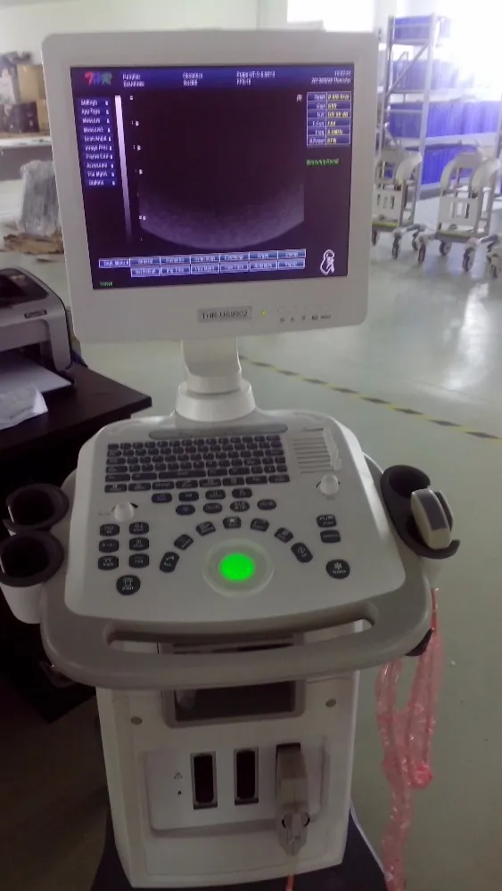
This high-precision Full Digital Image Technology system integrates advanced processing for superior clinical diagnostics.
DBF Digital Beam-forming & RDA Real-time Dynamic Aperture for crisp clarity.
Supports B, 2B, B/M, M, and 4B modes with THI Tissue Harmonic Imaging.
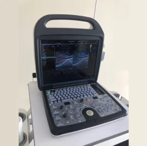
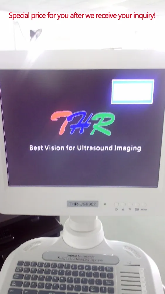
Windows XP Embedded system with one-key image optimization.
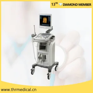
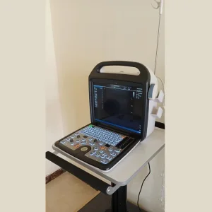
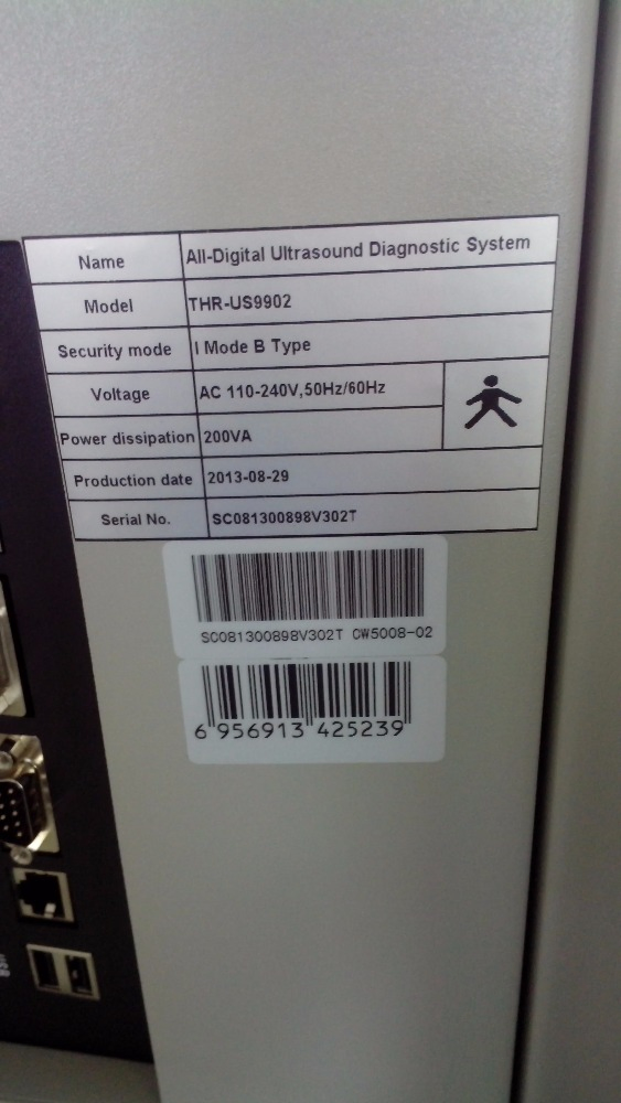
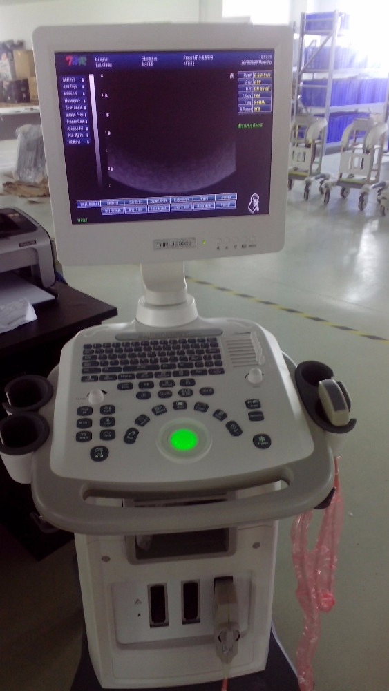
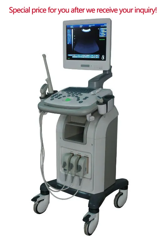
| Standard Unit | 15" LCD monitor, 3 Activated Sockets, Windows XP System |
| Adjustments | Smart 8 segments TGC adjustment |
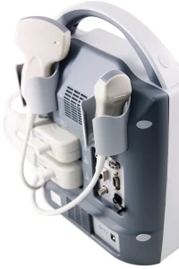
| Connectivity | PAL-D, VGA, RS-232, USB 2.0, DICOM 3.0 |
| Probe Options | Convex, Transvaginal, Linear, Micro-Convex, Rectum Linear |
| Keyboard | Backlight silica gel PBT keyboard for darkroom operation |
| Advanced Port | Video port, S-Video, RS-232, USB 2.0, VGA, DICOM 3.0 |